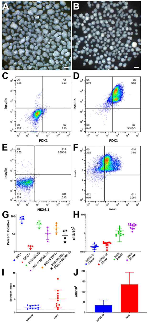
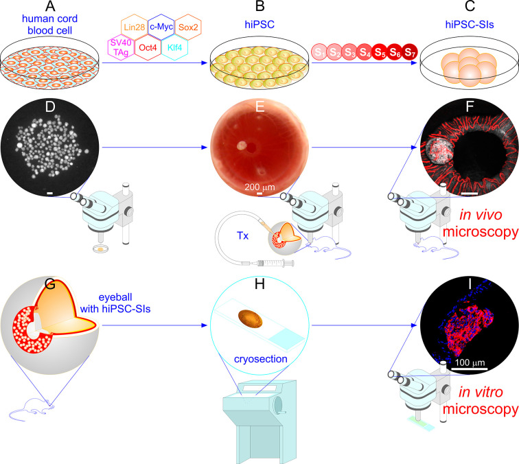
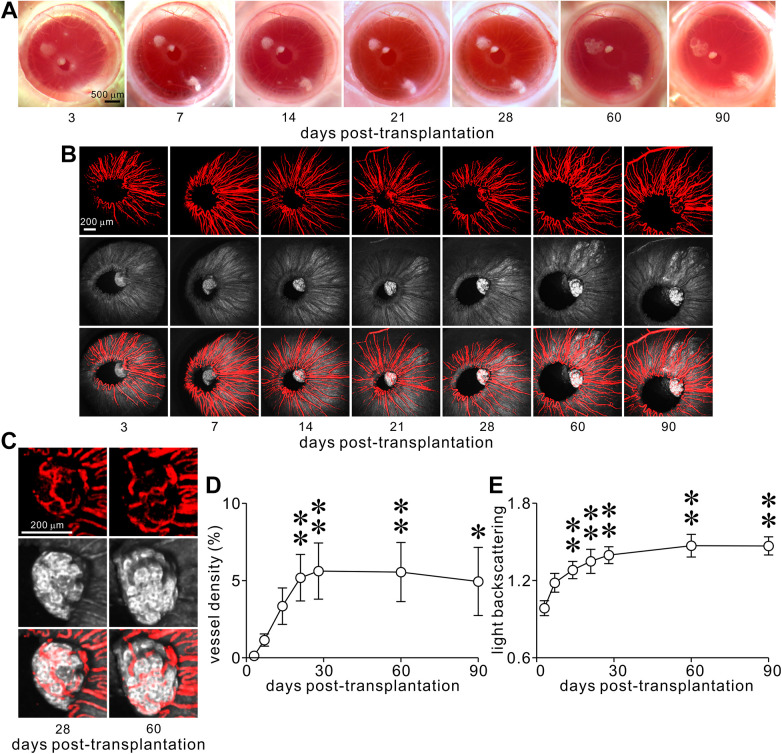
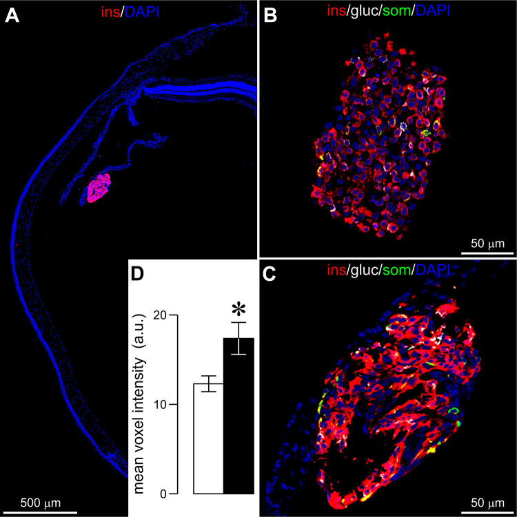
We exploited the anterior chamber of the eye (ACE) of immunodeficient mice as an ectopic site for both transplantation and microimaging of engineered surrogate islets from human induced pluripotent stem cells (hiPSC-SIs). These islets contained a majority of insulin-expressing cells, positive or negative for PDX1 and NKX6.1, and a minority of glucagon- or somatostatin-positive cells. Single, non-aggregated hiPSC-SIs were satisfactorily engrafted onto the iris. They underwent gradual vascularization and progressively increased their light scattering signals, reflecting the abundance of zinc-insulin crystal packaged inside mature insulin secretory granules. Intracameral hiPSC-SIs retrieved from recipients showed enhanced insulin immunofluorescence in correlation with the parallel increase in overall vascularization and light backscattering during the post-transplantation period. This approach enables longitudinal, nondestructive and intravital microimaging of cell fates, engraftment, vascularization and mature insulin secretory granules of single hiPSC-SI grafts, and may offer a feasible and reliable means to screen compounds for promoting in vivo hiPSC-SI maturation.
Keywords: backscattering; human induced pluripotent stem cells (hiPSCs); in vivo confocal microscopy; surrogate islet; the anterior chamber of the eye (ACE); transplantation; vascularization.